Echogenic intracardiac focus refers a small bright spot seen in the fetal heart during ultrasound examination. The exact underlying reason is unknown. It is postulated to be due to deposition of minerals or calcium within the papillary muscles of the heart;
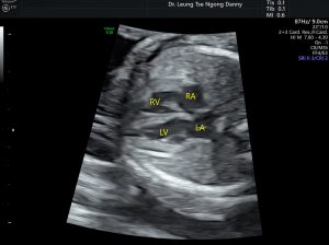
Normal fetal heart
It is present in 3-5% of normal fetuses on mid-trimester ultrasound and can be singular or multiple. Resolution is likely as the gestational age advances;
It is not associated with congenital cardiac malformation;
However, there is a weak association with Downs and Patau syndrome;
A fetal morphology scan is indicated. First, it is important to confirm the diagnosis and to exclude the rare possibility of cardiac tumour such as rhabdomyoma. Echogenic foci are usually smaller, < 3 mm in size while rhabdomyoma is usually bigger. Echogenic foci are found more commonly before 22 weeks of gestation while rhabdomyoma is usually found after 22 weeks of gestation. Rhabdomyoma might be associated with extracardiac lesions. However, these are not fast and fixed phenomena. In case of uncertainty, a follow-up ultrasound is useful for any increase in size, as expected in the case of cardiac tumour;
With the confirmation of the diagnosis on ultrasound, detailed search for any additional ultrasound markers for chromosomal abnormalities is needed.
For isolated echogenic intracardiac focus (i.e. no other ultrasound markers for chromosomal abnormalities), antenatal screening with OSCAR or NIPT test is considered adequate. An amniocentesis is not advised, unless the couple wants a 100% certainty on the fetal karyotype.
This article is contributed by Dr. T.N. Danny Leung