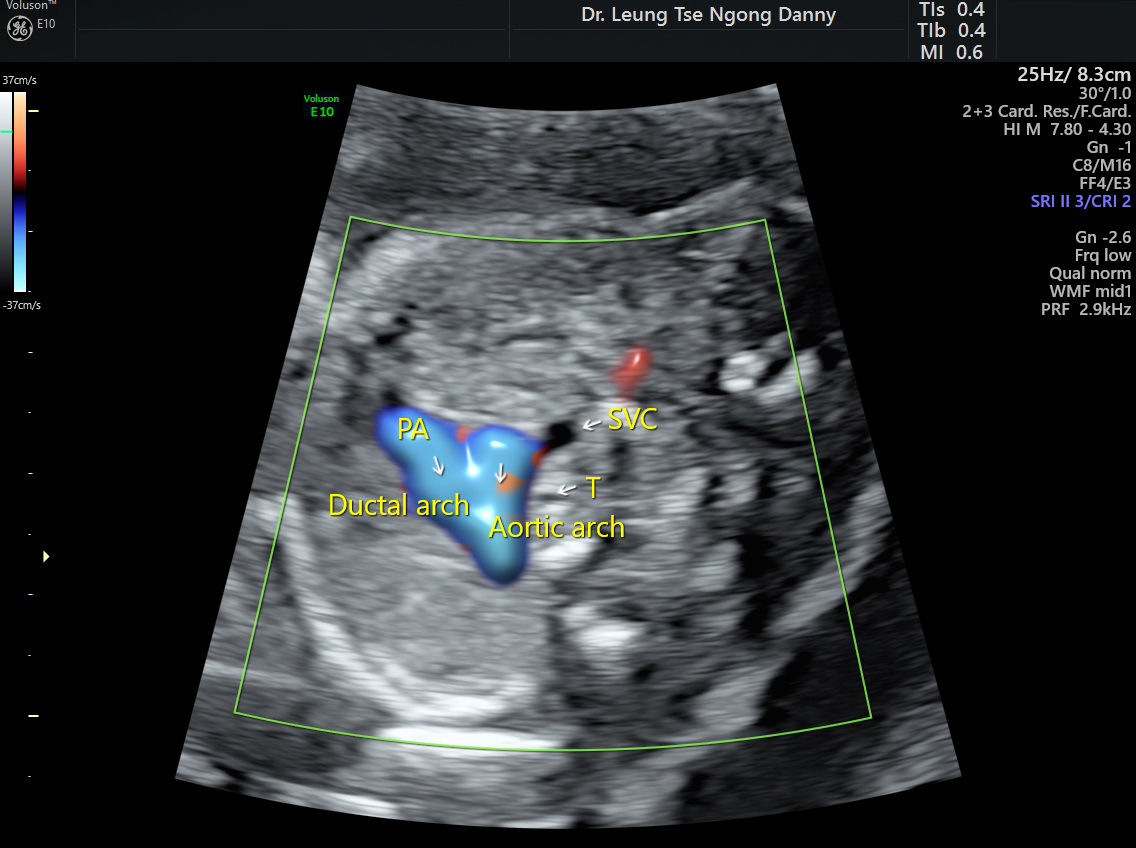
There are normally two major veins responsible for returning deoxygenated blood collected from the body back into the heart: the superior vena cava and the inferior vena cava. The superior vena cava (SVC) is responsible for venous blood from the head, neck and the upper limbs. It is located in the central front part of the thoracic cavity, to the right of the arch of aorta and to the front of the trachea. The SVC can be seen at the 3 vessels and trachea view during the fetal heart assessment in the second trimester;
Normal SVC in 3 vessels & trachea view
Occasionally the SVC is seen on the left side and it is called ‘persistent left SVC’. The term ‘persistent’ is used because the left SVC would have obliterated in early pregnancy and the failure of its involution results in its persistence. It happens in 0.3% of the population and is the most common form of venous malformation within the chest;
For persistent left SVC, a normal ‘right-sided’ SVC can be found in ~90% of cases, which is called ‘SVC duplication’;
It is associated with congenital heart abnormalities (5-9%) such as atrial septal defect, ventricular septal defect, coarctation of aorta and heterotaxy syndrome;
It is also a marker for chromosomal abnormalities, which was reported in ~12% of overall fetuses complicated by the condition and in 7% of isolated cases;
A detailed fetal morphology scan is indicated for ultrasound markers, fetal heart malformation and extra-cardiac lesions;