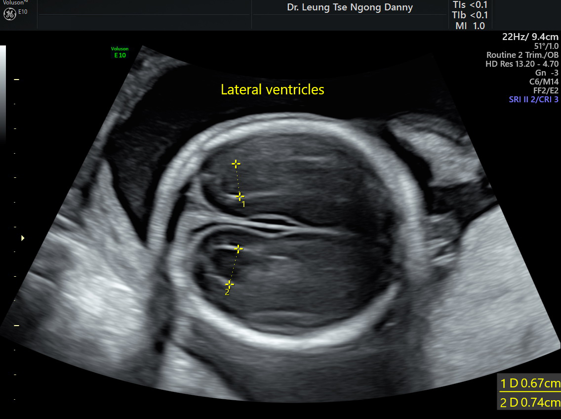
The average width of the lateral ventricle of the fetal brain on each side is 7.0 mm. The upper limit of normal is 10.0 mm. A thickness between 10.0- 15.0 mm is considered ‘borderline ventriculomegaly’ while a dimension thicker than 15.0 mm is diagnostic of hydrocephalus;
Normal lateral ventricles
Once ventriculomegaly is suspected, a detailed fetal morphology scan is recommended to look forother ultrasound markers or structural abnormalities. However, there is limitation of antenatal morphology scan as some abnormalities might not be picked up and the condition can be progressive. Also, antenatal ultrasound cannot predict the brain function in a long run;
There is an association with chromosomal anomalies (3% trisomy 21), Cytomegalovirus infection (< 2 %) and less commonly, some genetic syndromes. Amniocentesis for fetal karyotyping, array cGH and CMV PCR should be considered;
Counselling on the long-term prognosis for isolated borderline ventriculomegaly is known to be difficult. This ultrasound finding could well be a normal variant. In general, the long-term prognosis for isolated cases is good: normal neurodevelopment in 85% but in the remaining 15%, mild to severe psycho-motor or developmental delay have been reported. The prognosis has been shown to be better for size less than 12mm, absence of brain asymmetry and not being progressive in the later part of pregnancy;
MRI of the fetal brain is a new modality of imaging which might offer additional information on the fetal intracranial structures. Provided such service is available, it can be considered in this condition.
This article is contributed by Dr. T.N. Danny Leung