Club foot (also called congenital Talipes equinovarus) is a birth defect where the foot is rotated inward and downward. It can affect one (unilateral) or both (bilateral) feet and ~50% of babies have bilateral involvement;
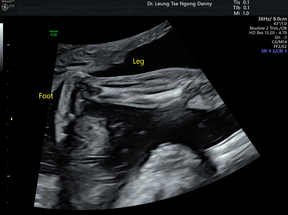
Normal leg & foot
The incidence is ~1 in 1,000 births;
It can be picked up on antenatal ultrasound. Occasionally the appearance of ‘club foot’ can be transient due to the fetal position. Oligohydramnios (reduced amniotic fluid volume), uterine anomalies and multiple pregnancy might lead to positional club foot. If the appearance of ‘club foot’ is persistent over a period of time, particularly even after the affected foot can move freely, it is likely to be a fixed deformity;
In the majority of babies with congenital club foot, it is an isolated abnormality and the long-term prognosis is good. With postnatal splinting +/- surgery, the baby should usually be able to walk with no problem. Antenatal referral to an Orthopaedic surgeon for parental counselling on the postnatal treatment options might be of value;
In ~20% cases, it is associated with other structural, neurological or syndromic anomalies. Therefore, a detailed fetal morphology scan should be arranged for other potential structural defects;
Amniocentesis for fetal karyotyping and array cGH helps to look for chromosomal and genetic aberrations. It is advised in the presence of additional structural abnormalities. In the cases of isolated club foot on ultrasound, the benefit of an amniocentesis has to be weighed balance against the small risk of miscarriage associated with the procedure, which is in the order of 1 in 200-300.
This article is contributed by Dr. T.N. Danny Leung