Echogenic bowel refers to the observation on ultrasound in the mid-trimester that the fetal bowel appears to be brighter than usual. A common criterion for the diagnosis of ‘echogenic’ is that the brightness is similar to or greater than that of adjacent bone;
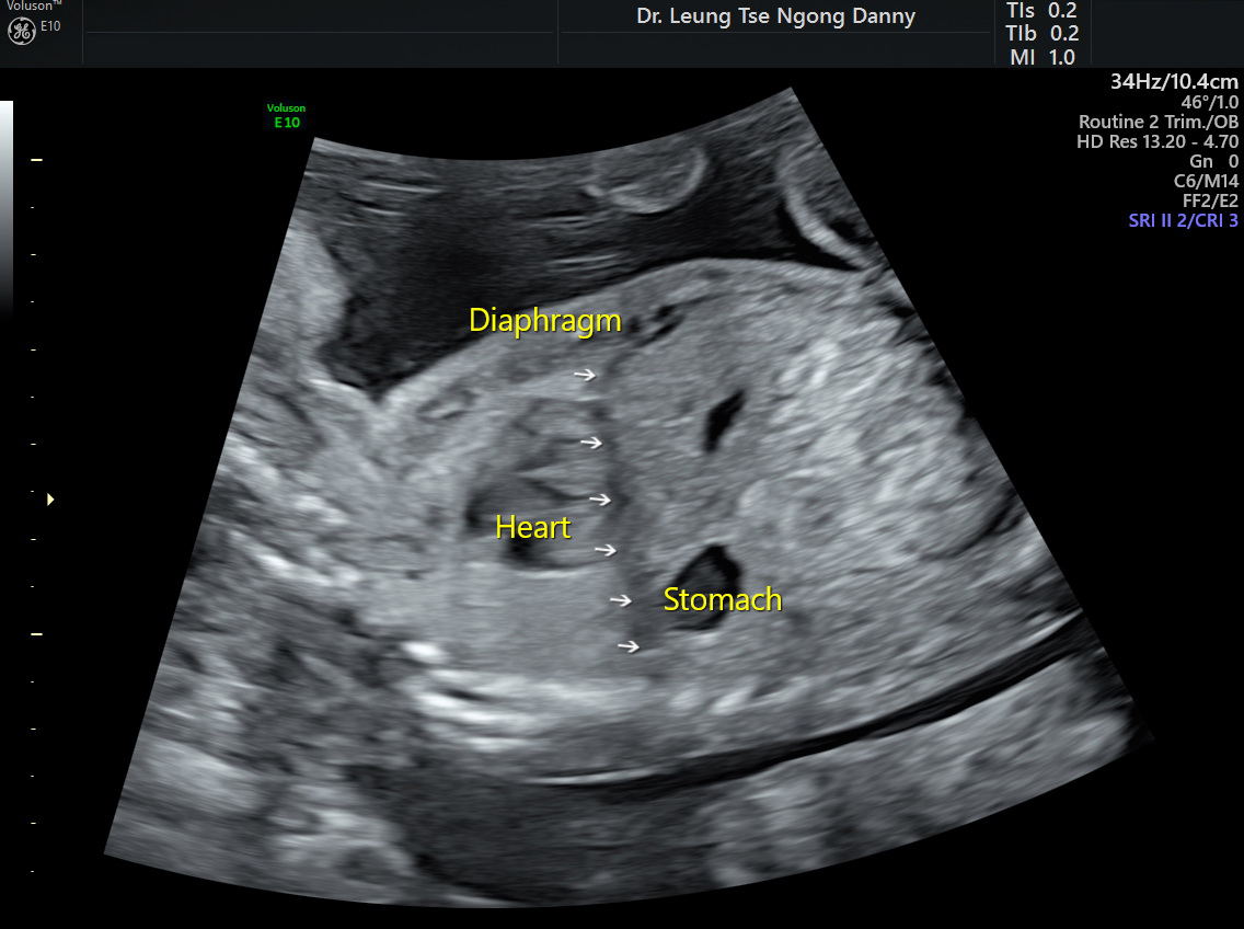
Normal abdomen & bowel
The transducer (the ultrasound probe) frequency used for scanning is important when considering the diagnosis of echogenic bowel. In one study, when using both 8 and 5 MHz transducers sequentially on the same fetuses, the diagnosis of echogenic bowel was made in 31% and 3% respectively. The use of high frequency transducer can lead to an over-diagnosis of the condition. Hence, for assessment of echogenic bowel, a transducer frequency of ≤5 MHz should be used;
Echogenic bowel can be found in normal fetus in up to 80-90% of cases. The echogenicity may resolve on a subsequent scan. The exact cause is unknown but one postulation is that intra-amniotic bleeding has occurred in early pregnancy, with subsequent swallowing of the blood into the fetal bowel;
It is also associated with pathological conditions such as chromosomal anomalies including trisomy 21, bowel abnormalities (including perforation or obstruction), cystic fibrosis, and Cytomegalovirus (CMV) infection;
A detailed fetal morphology scan is indicated to look for other ultrasound markers, bowel dilatation and evidence of CMV infection;
Maternal serological screening for CMV and Toxoplasma infection should be offered. For Caucacian parents, carrier screening for cystic fibrosis should be discussed. Cystic fibrosis is extremely rare among Chinese ethnic group;
For isolated echgenic bowel, it has been shown that the likelihood of having Downs syndrome is increased by 11 fold. The couple should be counselled regarding the limitation of morphology scan, the pros and limitation of NIPT test and the pros and risks of amniocentesis;
Follow-up scan is advised for monitoring the progress
This article is contributed by Dr. T.N. Danny Leung